
DLRLSS
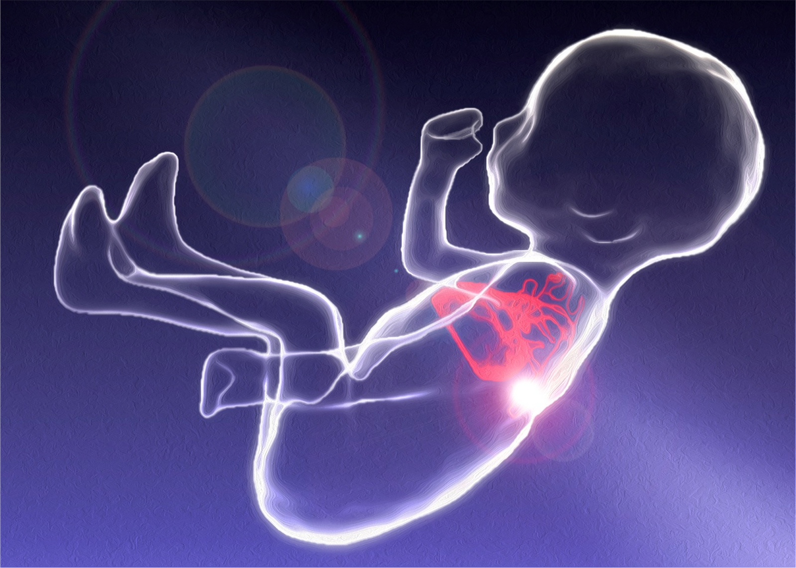
presentation on '3D/4D representation of fetal cardiac! anatomy and blood flow using MRI' for Deep Learning Reinforcement Learning Summer School at Alberta Machine Intelligence Institute
improved visualization and diagnosis of cardiac anomalies in utero by computational 3D ± time reconstruction from 2D MR images using unsupervised motion estimation, outlier detection and super-resolution to compensate for fetal-maternal move- ments and spatiotemporal limits of MR acquisition
Abstract
Magnetic resonance imaging (MRI) is a versatile imaging modality that is widely used to assess the morphology, function and tissue characteristics of the human heart. However, evaluation of the fetal heart relies principally on ultrasound, as fetal cardiac MRI is limited by the challenges associated with imaging a small, rapidly beating heart that is subject to various regular and spontaneous movements. These challenges are overcome by acquiring rapid 2D MR images, each providing limited view of the fetal heart in utero. The 2D images are then combined in a single 3D/4D reconstruction using unsupervised motion estimation, outlier detection and super-resolution imaging to compensate for fetal-maternal movements and spatio-temporal limits of MRI. This computational framework was applied to data collected in over 100 human fetal subjects, showing good agreement with echocardiography and improved diagnostic utility over 2D imaging for screening of structural cardiac anomalies in utero.
References
- Three-dimensional visualisation of the fetal heart using prenatal MRI with motion-corrected slice-volume registration. DFA Lloyd, K Pushparajah, JM Simpson, JFP van Amerom, MPM van Poppel, A Schulz, B Kainz, M Kuklisova Murgasova, M Lohezic, JM Allsop, S Mathur, H Bellsham-Revell, T Vigneswaran, M Charakida M, O Miller, V Zidere, G Sharland, MA Rutherford, JV Hajnal, R Razavi. The Lancet 393:1619-1627. doi: 10.1016/S0140-6736(18)32490-5.
- Fetal whole-heart 4D imaging using motion-corrected multi-planar real-time MRI JFP van Amerom, DFA Lloyd, M Deprez, AN Price, SJ Malik, K Pushparajah, MPM van Poppel, MA Rutherford, R Razavi, JV Hajnal. Magnetic Resonance in Medicine 82:1055-1072. doi: 10.1002/mrm.27798.
- Fetal whole-heart 4D flow cine MRI using multiple non-coplanar balanced SSFP stacks. TA Roberts, JFP van Amerom, A Uus, DFA Lloyd, AN Price, J-D Tournier, LH Jackson, SJ Malik, MPM van Poppel, K Pushparajah, MA Rutherford, R Rezavi, M Deprez, JV Hajnal. bioRxiv 2019:635797. doi: 10.1101/635797.
Videos
Contact
Joshua van Amerom
King's College London, School of Imaging Science & Biomedical Engineering (previous)
SickKids Hospital, Toronto, Division of Pediatric Cardiology (current)
joshua.vanamerom@sickkids.ca