
Intelligent Imaging 2015-05-14
Fetal Cardiac Cine Imaging Using Realtime MRI and Image-Based Spatiotemporal Motion Correction
14 May 2015
Joshua FP van Amerom
MRI is increasingly being used to image the fetus as an adjunct to ultrasound, however studies of the fetal heart and great vessels have found limited clinical utility [1]. Key challenges are fetal and maternal motion, a rapidly pulsating fetal heart, and extracting fetal ECG. Recent work has addressed the last point using self-consistency in reconstruction to infer a gating signal [2]. Here we take an approach based on realtime MR imaging and motion correction.
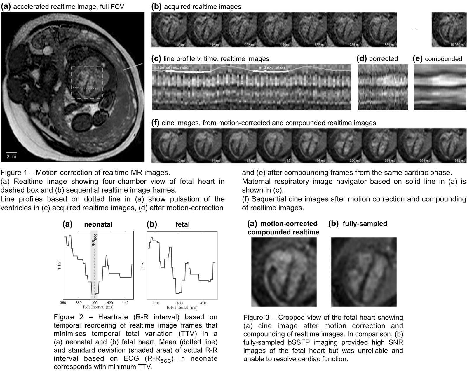
Realtime, full field-of-view

Realtime, cropped field-of-view

Cine from spatial+temporal motion correction of realtime, cropped field-of-view

Fetal singletons (28-37 weeks gestational age) were scanned in utero with a 1.5 T MRI system (Philips) using a 32-channel receiver coil in short and long axis cardiac views. Imaging was performed using a realtime dynamic 2D balanced steady-state free precession (bSSFP) sequence using partial-Fourier and parallel imaging to achieve a temporal resolution of 90-180 ms with a total under-sampling factor of 3-5 (TR/TE 4.4/2.2 ms, FA 60°, FOV 400 x 300-400 mm, voxels 1.8 x 2.3 x 5.0 mm). Realtime image series were processed as follows:
-
visual assessment to reject images in which the fetus was actively moving
-
image navigator to select frames at end expiration of the maternal breathing cycle
-
temporal reordering of image frames by minimising the temporal total variation (TTV) in the fetal heart between frames to identify the heart rate
-
spatial motion-correction using in-plane rigid registration of image frames
-
compounding images from the same cardiac phase to improve SNR
Fully sampled bSSFP images, with a temporal resolution of 573-765 ms, were acquired for comparison. Low SAR, < 2W/kg, and acoustic noise settings were adopted throughout, necessarily limiting scanner performance. Methods were validated on adult and neonatal cardiac data against ECG-gated cine sequences.
Figure 1 shows an example of motion compensation of realtime images. Figure 2 shows TTV plotted against heart rate for a neonatal case where ECG was also measured, and for a fetus. In all cases an unambiguous heart rate was detected. Compounded images preserved resolution while improving SNR and compared favourably to slower imaging, which was unreliable and unable to time-resolve cardiac function, as shown in Figure 3. Each step in the pipeline was tested individually and found to contribute to successful outcome.
A realtime imaging method of time resolved fetal cardiac imaging has been achieved. It can selectively incorporate non-contiguous sequences of images building robustness and tolerance to motion. There is potential for increasing both spatial and temporal resolution.
[1] Votino et al. US Obst Gyn 39(3)2012.
[2] Jansz et al. Mag Resn Med 64(5)2010.